


Glaucoma is a disease characterized by increased eye pressure and changes in the field of vision and the optic nerve. This disease is popularly known as green barbel. The normal level of eye pressure is from 12 to 22 mm Hg (mercury).
The origin and distribution of glaucoma :
The causes of increased eye pressure (intraocular pressure, hereinafter referred to as IOT) are different. We distinguish two basic groups of patients with regard to the width of the eye angle – with an open angle and with a closed angle. The second division of glaucoma divides them into those that arose primarily, that is, without a clear and proven reason, and those that arose secondarily as a result of some other eye disease. Here we will explain some of the most important forms, but first we need to explain how and where eye pressure occurs.
Aqueous fluid is produced in the ciliary body at a rate of 2-2.5 microliters per minute. From there it flows through the back chamber and the space between the lens and the iris into the front chamber. Then it enters the corner of the eye, where it is mostly resorbed, that is, it enters through the structures of the corner into tiny vessels that take it to the venous system of the eye and further into the bloodstream. If there is a disturbance in the ratio of formation and excretion of water, i.e. if, for any reason, more water is formed than it drains, an increase in eye pressure (intraocular pressure, IOT) occurs.
Chronic simple glaucoma (lat. Glaucoma chronicum simplex)
This type of glaucoma is caused by disturbances in the production and outflow of aqueous humor from the eye, but its exact cause has not been determined. With this type of glaucoma, we do not find mechanical changes in the eye that could be detected early and simply, but it looks completely normal until the first signs of increased eye pressure appear, which is usually after a long time. Namely, elevated eye pressure interferes with the circulation of the optic nerve and ganglion cells, causing their deterioration and weakening of function.
Angle-closure glaucoma
The disease occurs due to the closure of the angle by the iris, which “overlaps” the structure of the angle and mechanically prevents the outflow of aqueous humor. Then it accumulates in the eye and increases the pressure.
Secondary glaucoma (lat. Glaucoma secundarium)
It occurs for known reasons, i.e. an increase in IOT occurs due to other eye diseases. Here we will list the main causes:
Disorder of the eye lens
A thickened eye lens can press on the pupillary opening from the back and thus prevent the normal flow of fluid from the back chamber to the front. This situation most often occurs with some types of cataracts where the lens swells and causes a block. Injury to the eye lens by a foreign body causes its swelling and thus an increase in IOT.
The condition after cataract surgery
After cataract surgery, intraocular pressure increases relatively often due to the fact that remaining lens mass, remnants of liquid agents used during surgery, the position of the implanted artificial lens as well as blood elements change the spatial relationships in the eye and the possibility of fluid exiting through the structures of the corner of the eye. This can be treated very quickly and effectively with medication, so that such an increase in blood pressure returns to normal within one to two days and does not cause any consequences. However, in certain cases longer treatment is needed, and this is especially common in those people who had some types of glaucoma before surgery.
Eye inflammation
Eye inflammations (uveitis) are characterized by the presence of a large number of cellular elements in the eye chamber that are normally not present there or are present in only small amounts. These elements, as well as adhesions (synechiae) that are created between the iris and the lens or the cornea, mechanically prevent the normal flow of water as well as its resorption in the corner. With the withdrawal of the inflammation, the pressure normalizes, except in cases when the resulting growths remain in large numbers and continue to interfere with the natural circulation of the aqueous humor.
Diabetes
In advanced diabetic retinopathy, when new blood vessels are formed in places where there are normally not that many (iris and corner of the eye), the function of the corner and the resorption of the aqueous humor are weakened. This is especially complicated by additional bleeding in the eye, which is common in such advanced diabetes. The prognosis of the disease is poor and the patient is at risk of vision loss.
Eye injury
Mechanical, chemical and thermal injuries of the eye often affect its deeper structures. Despite operative or other appropriate treatment, they then irreversibly change the function of the eye and the relationships within it. The prognosis depends on the severity of the damage.
Congenital anomalies
Here we can talk about a wide range of changes in the structure of individual parts of the eye that are already visible from birth. It can be about disorders of the lens, iris, cornea or other parts of the eye. Fortunately, they are not common, and their treatment depends on each case.
Certain types of glaucoma often occur in several members of the same family. For this reason, after establishing the existence of glaucoma in one person, it is necessary to carry out an examination of other family members (siblings and children) in order to detect the possible existence of the disease in the earliest stage.
Symptoms:
Mild to moderate elevation of IOT is not easy to recognize by the patient because initially there are almost no symptoms. Only occasional mild pains and vague disturbances in the eye are possible. Only after a somewhat longer duration of glaucoma can weakening of vision occur due to the loss of function of the optic nerve. This happens because the elevated IOT adversely affects the circulation at the head of the optic nerve (papillas of the optic nerve, lat. Papilla nervi optici, PNO. Anatomically, it is the part of the optic nerve that is located in the eye itself), so due to its poor nutrition, it deteriorates . Equally elevated IOT also directly, mechanically destroys the optic nerve. Other ganglion nerve cells of the eye are affected by the same mechanism. With a stronger increase in IOT, fluid accumulates in the cornea (corneal edema), which blurs vision. When such a patient looks at the light around the light source (e.g. a light bulb) he sees luminous colored circles (halos) which are caused by the irregular scattering of light in the edematous cornea. This is a sign that the patient must see a doctor immediately, even if there are no other symptoms of the disease.
Acute glaucoma attack
A sudden and strong increase in IOT can occur in almost all types of glaucoma, but it is more common in angle-closure glaucoma and secondary glaucoma. The patient feels severe pain in the eye that spreads to the face and half of the head, and may also feel the urge to vomit. Vision is more weakened and halos are present. The eye is red and watery, and extremely hard to the touch. It is important to emphasize that an acute glaucoma attack is unilateral in most cases. If the symptoms are present on both sides, it is unregulated (decompensated) glaucoma that lasts for a long time. Attacks usually occur for no particular reason, except for angle-closure glaucoma, which occurs more often in dark rooms or movie theaters when the pupil dilates due to poor lighting and thus blocks the corner of the eye. A patient with the above-mentioned symptoms must report to the hospital immediately in order to stop the attack as quickly as possible and thus prevent consequences for the optic nerve.
Establishing a diagnosis:
Tests to prove glaucoma
In order to make a diagnosis of glaucoma, a whole series of tests is performed.
In Croatia, intraocular pressure (IOP) is measured mainly in two ways – the applanation method and Schiotz tonometry. In applanation tonometry, the eye is anesthetized with local anesthetic drops and fluorescein, a biological dye, is instilled. A specially designed prism is placed on the eye and the pressure level is read under cobalt blue lighting. Normal blood pressure is 12-22 mm Hg (mercury). With the Schiotz method, a tonometer is placed on the eye anesthetized with drops, on the scale of which the IOT is read. Normal measurement results using this method are from 5.5/4.0 to 5.5/7.0, where the first number indicates the weight of the weight used, and the second number indicates the height of the IOT. A higher second number means a lower IOT. For example, a pressure of 5.5/5.0 is normal, while a pressure of 5.5/2.0 is high. Other methods of measuring eye pressure are also used in some clinics.
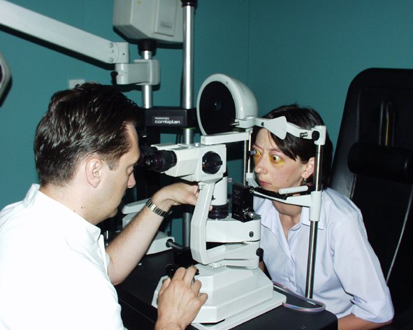
Applanation tonometry
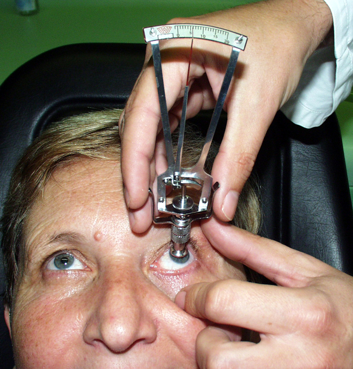
Tonometry according to Schiotz
Overview of the field of vision: the width of the field of vision means everything that can be encompassed (seen) when looking straight ahead with one eye. There are several basic ways of searching the field of view.
According to Goldmann, the visual field is an examination in which one looks at a standardized illuminated hemisphere, or at one specific black point in it. From time to time, small bright spots appear from the side, and the patient’s task is to give a sign as soon as he notices such a spot.
Computerized perimetry is a more modern and precise way of examining the field of vision, which can better determine the existence of certain outages in it. The viewing method can be compared to that of the field of view according to Goldmann, but the number of dots that appear is greater, and their appearance schedule is different, so the search itself can take quite a long time (several minutes to about 1 hour).
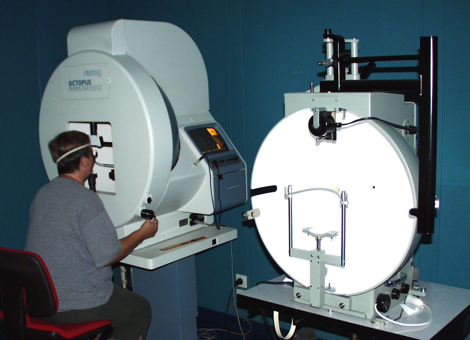
Computerized perimetry. In the front part, the device for examining the visual field according to Goldmann is visible.
Examination of the corner of the eye is part of the routine examinations that determine its width, which is necessary for the classification of the disease and its further treatment. With an open corner of the eye, the flow of the aqueous humor, i.e. its drainage path, is physically possible, but there are other obstacles that cause an increase in eye pressure. By a narrowed or closed angle of the eye, we assume a narrowing of the area of the eye angle due to the contact of the iris. This difference is fundamentally important and crucial for therapy, because in the case of a closed or narrowed angle, drugs should act on the iris in order to “pull” it out of the angle. This type of medication is called a miotic because it constricts the pupil.
The examination of the corner of the eye itself is performed with the help of a prism that contains mirrors placed at an angle. It rests on the anesthetized eye drops and thus enables visualization of the angle.
The examination of the background of the eye , among other things, tells about possible visible damage that occurred on the optic nerve itself. As already mentioned earlier, elevated IOT that lasts longer damages the optic nerve and it becomes pale and dies, and vision in that eye weakens. The damage caused in this way cannot be cured, but only slight improvements are possible, so it is necessary to treat the eye pressure on time and conscientiously so that it does not occur.
In addition to the above, a whole series of other tests are required to establish the diagnosis of glaucoma and to control the results of the treatment, i.e. its possible progression (examination of visual acuity, angle of the eye, etc.).
Treatment:
The goal of glaucoma treatment is to lower elevated IOT and prevent the consequences it can cause. In modern medicine, a large number of different eye drops and tablets are used that very effectively lower eye pressure. The mechanism of their action is to reduce the production of aqueous humor on the one hand and increase its outflow on the other. When using them, it is necessary to strictly follow the doctor’s instructions. Sometimes combinations of several types of drugs are needed. It can be said that in most cases the treatment of glaucoma is very long-term, and often lifelong. Regular check-ups by specialist doctors are required, and their number and frequency depends on the development of the disease. Apart from the above, the patient himself can do very little to lower IOT (sufferers often think that, for example, reading and watching television less often will protect their eyes). it is recommended to only avoid working in low light.Also, it should be known that elevated eye pressure is not related to elevated blood pressure, although the latter can also have consequences on the eyes.
Operative treatment of glaucoma is considered when it is not possible to effectively control eye pressure with other methods. It is performed in a hospital, and the type of procedure depends on the type of glaucoma. The goal of all types of surgery is to enable the aqueous humor to pass through alternative routes and bring it to a place where it can continue to flow naturally or be resorbed. In one of the most common anti-glaucoma operations, trepanotrabeculectomy, an opening is made in the sclera near the junction with the cornea, which does not heal, and which allows the passage of the aqueous humor and its arrival under the conjunctiva. This is where a so-called filtration bubble is created, from which water is resorbed, thus enabling the natural process of creation and outflow of aqueous humor.
Literature:
M.Sc. sc. Pavan Dr. Joško, “Eye Diseases”, Zagreb 2003.
